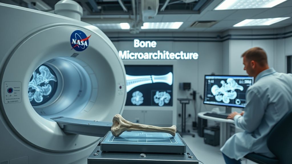
Introduction
Assessing bone quality goes beyond measuring bone density; it involves understanding how bone responds to mechanical stress, particularly repetitive or “cyclic” loading that can lead to fatigue failure. However, performing precise mechanical tests on the tiny bones of small animals like mice is extremely challenging, often leading to inconsistent data. This variability makes it difficult to detect subtle but important changes in bone quality caused by disease, aging, or environmental factors like spaceflight, especially when sample sizes are small. This study introduces a new, high-precision method to overcome these challenges and reliably measure the fatigue properties of mouse vertebrae.
Research Objective
The primary goals of this research were to:
- Develop a highly precise and repeatable ex vivo method for cyclic compressive loading of individual mouse vertebral bodies.
- Systematically reduce the experimental scatter in fatigue life measurements compared to standard techniques.
- Demonstrate the method’s superiority by comparing its results against two commonly used methods from existing literature.
Key Findings
The new method, termed the KFEA method, demonstrated significantly improved precision and accuracy over conventional approaches.
- The variability in the applied cyclic force was dramatically reduced, with a standard deviation 7- to 8-fold lower than the two literature-based methods (F-ratio = 5.5; p < 0.01).
- This precise force control led to a much more consistent measure of bone fatigue life, reducing the standard deviation by up to 5-fold compared to a conventional experimental method (F-ratio = 4.9; p < 0.01).
- Unlike the comparison methods, the KFEA method showed no correlation between applied force and fatigue life, confirming that all specimens were tested under a consistent initial strain.
- The use of a pivotable loading platen proved critical. A rigid platen, used in one comparison method, resulted in a 44.8% lower measured stiffness and a 40.9% shorter fatigue life, likely due to uneven loading and stress concentrations.
- The method produced highly uniform measurements for key bone quality parameters, including effective tissue elastic modulus (5.83 ± 0.22 GPa) and strain to failure (3.20 ± 0.80%).
Methodology
- Organisms Studied: The study utilized L5 vertebrae isolated from male C57BL/6J mice.
- Experimental Conditions: Each vertebral body was subjected to ex vivo cyclic compressive loading at 8 Hz in a saline bath until failure.
- Key Techniques: The novel method’s precision stems from a combination of three key innovations:
- 3D-Printed Jigs: Custom jigs were used to hold the tiny vertebrae securely, allowing for the machining of perfectly plano-parallel surfaces for uniform contact.
- Pivotable Loading Platen: A spherically-seated upper platen was used to self-align with the specimen’s surface, ensuring even load distribution and preventing localized stress.
- Micro-CT Finite Element Analysis (FEA): Before testing, each vertebra was scanned with micro-CT. A computational FEA model was created from the scan to calculate the specimen’s unique stiffness. This allowed researchers to prescribe a specimen-specific force that would produce the exact same target strain level in every sample, eliminating errors from machine compliance and inter-specimen variability.
Importance for Space Missions
Understanding how bone quality degrades during spaceflight is critical for ensuring astronaut health on long-duration missions to the Moon and Mars. This high-precision method offers a powerful tool for this research.
- Enhanced Detection Power: Spaceflight experiments are constrained by very small sample sizes. By minimizing data scatter, this method increases the statistical power to detect subtle but significant deterioration in bone’s fatigue resistance caused by microgravity and radiation.
- Evaluating Countermeasures: The technique provides a reliable platform to assess the effectiveness of countermeasures, such as new exercise regimens or therapeutic drugs, in preserving the mechanical integrity and fatigue resistance of bone.
- Beyond Bone Density: This method allows researchers to investigate changes in bone quality that are not captured by standard bone density (DXA) scans, providing a more complete picture of skeletal health and fracture risk.
Knowledge Gaps & Future Research
While this method represents a significant technological advance, it also highlights areas for future investigation:
- The protocol needs to be adapted and validated for other animal models (e.g., rats) and different types of bone (e.g., long bones).
- Further research is needed to establish a direct link between these ex vivo fatigue measurements and the actual risk of bone fracture in vivo.
- The next crucial step is to apply this method to analyze bone samples from animals flown on space missions to quantify the true impact of the space environment on bone quality.
- The method could be used to help distinguish the separate and combined effects of microgravity and space radiation on bone’s mechanical properties.
Results
This study successfully developed and validated a robust methodology that significantly improves the precision of biomechanical testing for small-animal bones. By integrating 3D-printing, advanced micro-CT imaging, and specimen-specific computational modeling, this technique provides a highly reliable platform for assessing bone fatigue life. Its ability to generate consistent data from small sample sizes makes it an invaluable tool for future space bioscience research, enabling a deeper understanding of how to protect astronaut skeletal health during extended missions.